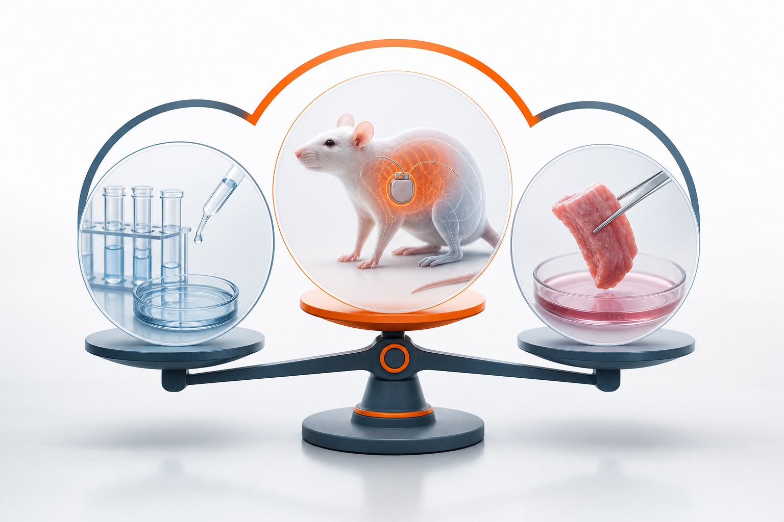
- Defining the Terms: In Vivo, Ex Vivo, and In Vitro
- In Vivo Testing in Medical Device Development
- Ex Vivo Testing in Medical Device Development
- How In Vivo and Ex Vivo Data Fit Into the Regulatory Submission
- Where Ex Vivo Testing Ends and In Vivo Testing Begins
- Practical Implications for First-in-Human Program Design
- In Vivo vs Ex Vivo in Radiopharmaceutical Trials
- Choosing the Right Testing Sequence for Your Device
- Frequently Asked Questions
Medical device sponsors mapping their preclinical-to-clinical pathway encounter the in vivo vs ex vivo distinction early and often. The choice between these two testing approaches shapes protocol design, regulatory documentation, and the evidence package you will eventually submit to the FDA. Getting the terminology right matters before you write a single line of your Pre-Submission meeting request.
This article explains what each method means, how they differ from in vitro work, where each fits in the device development sequence, and what the distinction means for your first-in-human (FIH) program.
Defining the Terms: In Vivo, Ex Vivo, and In Vitro
The three Latin prefixes do the heavy lifting here.
In vivo means "within the living." Testing is performed inside a living organism — an animal model during preclinical work, or a human subject during a clinical trial. The device, drug, or intervention interacts with a complete, functioning biological system in real time.
Ex vivo means "outside the living." Biological material — tissue, an organ, or cells — is removed from a living organism and kept viable in a controlled environment. The device is then tested against that material outside the body. The tissue is real and biologically active, but the systemic context of a whole living organism is absent.
In vitro means "within the glass." Testing occurs entirely in an artificial environment: a cell culture, a bench-top fluid model, or a synthetic tissue analog. No living organism is involved.
These three methods are not interchangeable. Each answers a different class of question, and each carries a different weight in a regulatory submission.
In Vivo Testing in Medical Device Development
In vivo testing is the gold standard for understanding how a device behaves inside a complete physiological environment. Blood flow dynamics, immune response, tissue remodeling, and systemic tolerability can only be observed when the full biological system is present and active.
In preclinical programs, in vivo studies typically use animal models selected for anatomical or physiological similarity to the human target. A cardiovascular device might be evaluated in a porcine model because swine coronary anatomy closely resembles human anatomy. A spinal implant might use an ovine or caprine model for bone density comparability.
The outputs of a well-designed in vivo animal study feed directly into the IDE application and the Pre-Submission package. FDA reviewers expect in vivo biocompatibility data, fatigue performance under physiological loading, and — for implantable devices — histopathology from explanted tissue. The ISO 10993 series governs the biological evaluation framework; ISO 14155 governs clinical trial conduct once you cross into human subjects.
In vivo human studies, meaning clinical trials, are the endpoint of the entire development sequence. The first-in-human study is the first in vivo human test of the device. That distinction is worth stating plainly: every prior in vivo test, however rigorous, was a model. The FIH study is the real thing.
Ex Vivo Testing in Medical Device Development
Ex vivo testing occupies a useful middle ground. It is more physiologically relevant than in vitro bench work because the tissue or organ retains its biological structure and cellular activity. It is also less complex than a full animal study — no systemic immune response, no circulatory pressure variation, no anesthesia protocol to manage.
Common ex vivo applications in device development include:
- Valve and vascular testing: Excised porcine or bovine hearts are perfused with fluid to simulate cardiac conditions. A transcatheter valve or endovascular device can be deployed and evaluated under realistic tissue conditions without a live animal study.
- Soft tissue and wound closure: Excised skin or muscle tissue allows evaluation of suture pull-out strength, staple integrity, or adhesive performance against real biological material.
- Orthopedic and spinal implants: Cadaveric bone specimens provide realistic bone density, cortical thickness, and trabecular structure that synthetic bone analogs cannot replicate.
- Ophthalmic devices: Excised porcine or bovine eyes are used extensively to test intraocular lenses, surgical instruments, and drug delivery systems before animal or human studies.
- Radiopharmaceutical dosimetry: Ex vivo tissue samples can be used to measure radiolabeled compound uptake and retention at the cellular level before proceeding to in vivo dosimetry studies.
The regulatory value of ex vivo data depends on how well the model is characterized. FDA and notified bodies will scrutinize the tissue source, the preservation method, the temperature and perfusion conditions, and the duration of the test. An ex vivo study that is poorly documented provides limited regulatory credit even when the results are favorable.
How In Vivo and Ex Vivo Data Fit Into the Regulatory Submission
FDA's framework for IDE applications under 21 CFR Part 812 requires a body of preclinical evidence sufficient to support the safety of the proposed human study. That evidence package typically combines in vitro bench data, ex vivo functional testing, and in vivo animal data. The weight given to each depends on the device classification, the intended use, the implant duration, and the contact type.
For a significant-risk device pursuing an IDE, the preclinical section of the submission must demonstrate that the device performs as intended under conditions that approximate clinical use. In vivo animal data carries the most weight because it reflects systemic response. Ex vivo data supports specific performance claims — particularly for mechanical or functional endpoints that are difficult to isolate in a live animal. In vitro data establishes baseline material and design characterization.
Data collected under ISO 14155 protocol architecture and structured per FDA 21 CFR 812.28 is accepted for US IDE and IND submissions when the clinical study is conducted outside the United States. That pathway is directly relevant to sponsors who conduct their FIH study in Latin America and intend to use that data to support a US pivotal trial or marketing submission.
The Cook Group's multi-site first-in-human artificial venous valve study — managed by bioaccess® in Colombia, with more than 142 INVIMA regulatory submissions handled — is one example of how in vivo human data generated outside the US feeds a US regulatory strategy. Program details are available in the Cook Advanced Technologies first-in-human venous valve case study.
Where Ex Vivo Testing Ends and In Vivo Testing Begins
The boundary between ex vivo and in vivo is not always obvious in practice. A few clarifying scenarios help.
A device tested in an excised porcine heart on a bench — even one being actively perfused — is ex vivo. The moment that same device is implanted in a living pig under anesthesia, the study is in vivo. A cell culture assay using primary human cells derived from a biopsy is ex vivo at the tissue level but in vitro at the study-design level. The distinction matters for how you categorize the study in your regulatory submission and how you describe it in your clinical investigation plan.
For gene therapy and cell therapy products, the ex vivo/in vivo distinction carries additional regulatory weight. Ex vivo gene modification involves removing cells from a patient, modifying them outside the body, and reinfusing them. In vivo gene delivery introduces the vector directly into the patient. Medical device sponsors working on combination products that include a biological component — such as a scaffold seeded with autologous cells — need to address both modes in their preclinical package.
The Envveno Medical program, which pursued the first-ever FDA IDE for a non-surgical replacement venous valve, required a preclinical evidence package addressing device performance across multiple testing modalities before proceeding to human subjects. The Envveno Medical LATAM FIH case study illustrates how that evidence sequence translates into a submission-ready package.
Practical Implications for First-in-Human Program Design
When building the preclinical plan that will support your IDE application and FIH protocol, the in vivo vs ex vivo distinction has direct scheduling and budget implications.
Ex vivo studies are generally faster and less expensive than in vivo animal studies. They do not require IACUC approval, do not involve animal husbandry costs, and can often be completed at a contract testing laboratory on a shorter timeline. For sponsors managing financial runway carefully, front-loading ex vivo work to de-risk the design before committing to a GLP animal study is a sound strategy.
In vivo animal studies take longer to plan, execute, and report. GLP compliance, histopathology processing, and veterinary oversight all add time. But they are not optional for most significant-risk implantable devices. FDA expects in vivo data for implant duration, tissue response, and systemic tolerability — endpoints that ex vivo models cannot provide.
The ClarVista Medical program, which proceeded from first-in-human to an Alcon acquisition, demonstrates how a well-sequenced preclinical and clinical evidence strategy supports both regulatory approval and commercial outcomes. The ClarVista Medical LATAM success story provides a concrete reference for how that sequence plays out.
Once in vivo animal data supports the safety case, the FIH study is the next in vivo step. The testing environment shifts from an animal model to a human subject, and the regulatory framework shifts from preclinical standards to ISO 14155 and the IDE regulations.
In Vivo vs Ex Vivo in Radiopharmaceutical Trials
The distinction carries specific meaning for radiopharmaceutical developers working with Lu-177, Ac-225, or Ga-68 compounds. Ex vivo biodistribution studies — where a radiolabeled compound is administered to an animal and tissues are harvested and counted after sacrifice — are a standard preclinical method for characterizing tissue uptake and clearance. These studies are technically in vivo at the administration stage and ex vivo at the measurement stage. Regulatory submissions for radiopharmaceutical INDs typically distinguish between the two phases explicitly.
In vivo imaging studies, such as PET or SPECT scans in animal models or human subjects, measure biodistribution without tissue harvest. These are fully in vivo and provide dynamic data that ex vivo counting cannot replicate.
For sponsors developing targeted radiopharmaceuticals, the preclinical package typically includes both ex vivo biodistribution data and in vivo imaging data before proceeding to a Phase 0 or Phase I human study. The CelonOva BioSciences COBRA PzF coronary stent program, which involved polymer-free drug elution evaluation, required a similarly layered preclinical evidence approach. The CelonOva BioSciences case study reflects how that evidence architecture supports a submission-ready package.
Choosing the Right Testing Sequence for Your Device
No single testing sequence fits every device type. The right approach depends on your device classification, the intended patient population, the implant duration, the contact type, and the specific safety questions your IDE reviewer will need answered.
A general framework for early-stage device sponsors:
- In vitro bench testing establishes design characterization, material properties, and dimensional verification.
- Ex vivo functional testing evaluates performance under realistic tissue conditions and surfaces design issues before animal studies.
- In vivo animal studies under GLP conditions demonstrate biocompatibility, systemic tolerability, and implant-duration response.
- First-in-human clinical study, conducted under ISO 14155 and structured per FDA 21 CFR 812.28, generates the human safety and performance data that supports the IDE and subsequent pivotal trial.
Each stage informs the next. Gaps in the sequence create questions that FDA reviewers will surface in their Pre-Submission response or IDE review letter — and addressing those gaps before submission is far less costly than responding to a deficiency letter after the fact.
If your program is approaching the FIH stage and you need a submission-ready evidence package within a defined timeline, bioaccess® structures that process through the FIH-12™ program. Learn more at bioaccessla.com.
Frequently Asked Questions
What is the difference between in vivo and ex vivo testing in medical devices?
In vivo testing is performed inside a living organism, capturing the full systemic biological response. Ex vivo testing uses biological material removed from a living organism and kept viable outside the body — more physiologically relevant than synthetic bench models, but without the complexity of a full animal study.
Is ex vivo testing considered preclinical or clinical?
Ex vivo testing is preclinical. It does not involve human subjects and does not require an IDE or IND. It generates supporting evidence for the preclinical section of a regulatory submission, not clinical data under ISO 14155 or FDA 21 CFR Part 812.
Can ex vivo data replace in vivo animal studies in an IDE submission?
Generally, no. FDA expects in vivo animal data for significant-risk implantable devices to address systemic tolerability, tissue response, and implant-duration endpoints that ex vivo models cannot replicate. Ex vivo data supports specific functional or mechanical performance claims but does not substitute for GLP in vivo studies.
How does in vivo data from a Latin American FIH study support a US IDE?
Data collected under ISO 14155 protocol architecture and structured per FDA 21 CFR 812.28 is accepted for US IDE and IND submissions. A first-in-human study conducted in Panama, Colombia, Chile, or another Latin American country under that framework generates human safety and performance data that FDA will accept as part of a US submission package.
What is the difference between in vivo and in vitro testing?
In vivo testing occurs inside a living organism. In vitro testing occurs entirely in an artificial environment — a cell culture or bench-top fluid model — with no living organism involved. In vitro data is the least physiologically representative of the three methods but is the fastest and least expensive to generate.
When should a device sponsor run ex vivo studies versus proceeding directly to animal studies?
Ex vivo studies are most useful when you need to evaluate device performance against real biological tissue before committing to the cost and timeline of a GLP animal study. They are particularly valuable for vascular, cardiac, orthopedic, and ophthalmic devices where tissue interaction is central to the performance claim. They are not a substitute for in vivo animal studies when systemic response data is required.
What regulatory standards govern in vivo clinical testing of medical devices?
ISO 14155 governs the conduct of clinical investigations of medical devices in human subjects. FDA 21 CFR Part 812 governs IDE applications and the conduct of investigational device studies in the United States. For foreign clinical data to be accepted in a US submission, the study must be structured per FDA 21 CFR 812.28.
Understanding the in vivo vs ex vivo distinction is foundational work. It shapes how you design your preclinical package, how you sequence your development program, and how you frame the evidence in your IDE submission. Getting that foundation right before you engage with FDA reviewers or select a CRO saves time that most early-stage sponsors cannot afford to lose.
Leave a Reply